Why Is Precise Diagnosis Crucial Before Selecting a Urinary Stone Surgery Hospital near Bongmyeong Station?
What makes urinary stones so painful, and why is prompt evaluation essential?
Urinary tract calculi are solid crystalline aggregates formed from mineral deposition within the renal pelvis, calyces, or ureter; as a progressive disease, they can migrate to obstruct urine flow, leading to severe acute renal colic, hydronephrosis, and potentially life-threatening urosepsis. When a stone blocks the narrow lumen of the ureter, the proximal urinary tract spasms and distends, triggering some of the most intense physical pain known in clinical medicine. Many residents searching for a specialized urology clinic near Bongmyeong Station are driven by this sudden, excruciating pain to seek immediate surgical options. However, clinical urgency should always be balanced with an accurate diagnostic protocol to prevent unnecessary invasive procedures.
Immediate medical evaluation is crucial because prolonged obstruction can cause irreversible damage to the renal parenchyma. Within 48 hours of complete obstruction, renal blood flow begins to decline, and permanent nephron loss can start if the blockage is not resolved within one to two weeks. Therefore, the primary goal of visiting a urology clinic is not just pain relief, but a comprehensive assessment of renal function and stone morphology to determine whether conservative management is safe or if surgical intervention is mandatory.
Treatment timing: Active intervention is urgently required if the stone exceeds 10mm, causes unmanageable pain, or is accompanied by signs of infection such as fever and chills.
Non-surgical care: Conservative management with high-volume hydration and medical expulsive therapy is highly reasonable for uncomplicated stones under 5mm located in the distal ureter.
Treatment selection: The choice between Extracorporeal Shock Wave Lithotripsy (ESWL) and retrograde intrarenal surgery (RIRS) depends on stone density (measured in Hounsfield Units via CT), anatomical features of the renal calyx, and the patient’s bleeding risks.
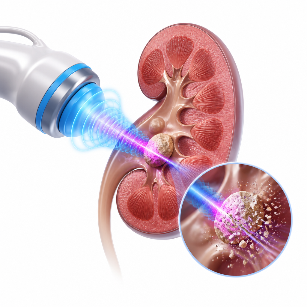
How do clinical guidelines distinguish between conservative management and active surgical intervention?
According to domestic and international clinical guidelines, including those published by the European Association of Urology (EAU) in 2023, the management of urinary stones is strictly categorized based on objective quantitative criteria. For stones smaller than 5mm without anatomical anomalies or signs of urinary tract infection, conservative management is considered the gold standard. This non-surgical approach primarily utilizes medical expulsive therapy (MET) featuring alpha-blockers (such as tamsulosin) to relax the smooth muscle of the distal ureter, combined with oral hydration of at least 2.5 to 3 liters daily to facilitate spontaneous passage.
In contrast, active intervention becomes necessary when the stone is larger than 10mm, as the probability of spontaneous expulsion drops below 20%. For stones sized between 5mm and 10mm, the decision-making process is more nuanced. Factors such as persistent hematuria, recurrent renal colic, patient occupation (e.g., pilots or frequent travelers), and signs of progressive hydronephrosis must be thoroughly analyzed. Furthermore, if a patient presents with a fever exceeding 38°C alongside flank pain, it suggests obstructive pyelonephritis, which constitutes a medical emergency requiring immediate drainage via a ureteral stent or percutaneous nephrostomy before any lithotripsy is attempted.
| Treatment Option | Primary Indications | Key Advantages | Clinical Limitations |
|---|---|---|---|
| Conservative Care (MET) | Stones < 5mm, minimal pain, no infection | No anesthesia, zero procedural risks, low cost | Unpredictable timeline, risk of sudden severe pain |
| ESWL (Shockwave) | Stones < 15mm, low density, high renal location | Outpatient procedure, no incision or endoscopy | Lower success on hard stones (high Hounsfield Units) |
| RIRS (Flexible Endoscopy) | Stones > 15mm, lower pole calyx, hard composition | High stone-free rate (>90%), direct visualization | Requires general spinal anesthesia and ureteral stent |
According to clinical evidence compiled by the American Urological Association, both the physical dimensions of the stone and the patient’s individual anatomical structures must be evaluated using non-contrast computed tomography (NCCT) to optimize the safety of surgical interventions.

What is the key checklist for patients seeking a specialized urology clinic near Bongmyeong Station?
When selecting a urinary stone surgery hospital, patients in the Bongmyeong Station area should evaluate the clinical infrastructure of the urology department. A reliable clinic should possess state-of-the-art diagnostic equipment, such as low-dose digital X-ray and high-resolution ultrasound, and preferably a non-contrast CT scanner to accurately map the stone’s 3D coordinates and density. Additionally, the availability of both non-invasive treatment modalities like Extracorporeal Shock Wave Lithotripsy (ESWL) and advanced endoscopic options like Retrograde Intrarenal Surgery (RIRS) ensures that treatment is tailored specifically to the patient’s pathology rather than the clinic’s hardware limitations.
However, outcomes may differ in exceptional cases such as patients with anatomical anomalies like a horseshoe kidney, solitary kidneys, severe infundibulopelvic angles, or uncorrected bleeding diatheses where standard shockwave protocols cannot be safely applied. In these complex scenarios, referral to a tertiary medical facility or a highly specialized surgical center may be necessary to ensure safety.
Essential decision checklist for patients:
- Does the clinic provide 24/7 emergency medical response for sudden renal colic?
- Are both ESWL and endoscopic laser lithotripsy (RIRS/URS) available on-site?
- Is there a detailed diagnostic protocol, including Hounsfield Unit (stone density) evaluation?
- Are the specialists experienced in treating recurrent stones and managing postoperative double-J stents?
- Does the medical team offer personalized metabolic evaluation to prevent future recurrence?
If-Then Decision Flowchart for Urinary Stone Management:
- Step 1: If the stone is less than 5mm and the patient’s pain is well-controlled without systemic fever, then initiate conservative medical expulsive therapy (MET) with increased hydration for 2 to 4 weeks.
- Step 2: If the stone is between 5mm and 15mm with low density, then recommend Extracorporeal Shock Wave Lithotripsy (ESWL) as a primary, non-invasive outpatient treatment.
- Step 3: If the stone is larger than 15mm, highly dense, located in the lower renal pole, or if shockwave therapy has failed, then recommend Retrograde Intrarenal Surgery (RIRS) using flexible endoscopy and holmium laser.
Frequently Asked Questions FAQ
QIs shockwave lithotripsy (ESWL) always the best option for urinary stones?
No, ESWL is highly effective for moderate-sized stones in the upper urinary tract, but its success rate drops significantly for stones with high mineral density (greater than 1,000 Hounsfield Units on CT) or those located in the lower pole of the kidney where gravity hinders the clearance of broken fragments. In such cases, flexible endoscopic surgery (RIRS) is often preferred near Bongmyeong Station.
QHow long does the recovery take after undergoing RIRS endoscopic surgery?
RIRS is a minimally invasive endoscopic procedure, meaning patients typically return to light daily activities within 2 to 3 days. However, a temporary double-J ureteral stent is usually placed to prevent swelling, which may cause minor bladder irritation or hematuria during urination for 1 to 2 weeks until it is removed.
QWhy do urinary stones recur so frequently, and how can I prevent them?
Urinary stones have a 50% recurrence rate within five to ten years if lifestyle changes are not implemented. Preventive measures include drinking enough water to maintain a daily urine output of 2.5 liters, reducing sodium and animal protein intake, and maintaining dietary calcium levels to bind oxalates in the digestive tract before they reach the kidneys.
This content is general medical information, and individual treatment decisions should be made through imaging tests and in-person medical evaluation.
Author: Medical content editor based on medical information research
Reviewed by: Specialist consultation from the relevant department
Last reviewed: 2026-08-03
Reference guideline: 2023 European Association of Urology (EAU) Guidelines on Urolithiasis
Medical neutrality and closing note
The core of medical decision-making is not to follow a specific device or a trending procedure, but to choose an option that fits each patient’s individual anatomy, condition, risk level, and treatment goals. Every procedure has both advantages and limitations, so decisions should be made after sufficient discussion with an experienced specialist.
[Medical information and copyright notice]
This content is a professional medical column prepared based on medical consultation from 굿모닝비뇨기과.
The infographics used in this article are created to support understanding and may differ from actual clinical results.
The information provided is a general medical guideline, and accurate diagnosis and treatment require an in-person evaluation by a qualified specialist.