Introduction to Advanced Urological Screening
For international patients seeking specialized urological care, understanding the nuances of diagnostic protocols is the first step toward effective health management. Bladder cancer remains a significant concern globally, and patients traveling to medical hubs—particularly those exploring options in internationally recognized medical hubs like South Korea—often face complex questions regarding the accuracy and comfort of screening procedures. The focus of modern urology has shifted from simple detection to high-precision characterization of mucosal lesions, utilizing advanced imaging and molecular markers to ensure early intervention. Choosing a medical institution that meets these criteria is crucial for those who require definitive answers regarding their urological health.
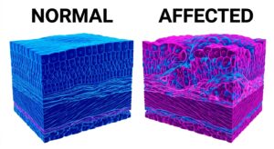
• Medically, bladder cancer is defined as a malignancy originating in the urothelium, the epithelial lining of the urinary bladder, characterized by the abnormal proliferation of cells that may present as painless gross hematuria or microscopic bleeding.
• Conditions under which conservative management or simple observation is reasonable include asymptomatic patients without hematuria or specific risk factors (such as heavy smoking history or occupational chemical exposure) who do not meet clinical screening thresholds.
• Criteria to consider when choosing treatment or screening include the sensitivity of the diagnostic modality (such as NBI cystoscopy), the patient’s anatomical risk profile, and the availability of a comprehensive recovery timeline for international travelers.
According to multiple observational studies and meta-analyses, early-stage bladder cancer detection significantly improves the 5-year survival rate, which can exceed 90% when the disease is confined to the inner lining of the bladder. However, in exceptional cases where the tumor has already invaded the muscularis propria, the clinical approach must pivot from screening to aggressive staging and multi-modal therapy. South Korea has developed standardized protocols based on extensive case volume, allowing for high-throughput yet personalized diagnostic pathways that cater to both local and international populations.

The Pathophysiology of Urothelial Malignancy
The fundamental principles of bladder cancer relate to the unique environment of the urinary tract. The urothelium acts as a barrier against toxic metabolites in urine, but chronic exposure to carcinogens can trigger genetic mutations in the fibroblast growth factor receptor (FGFR) or the TP53 gene. Clinical data from Korean medical centers suggests that the speed of technology adoption in these facilities allows for the integration of genetic sequencing and advanced cytology into standard screening visits. This is particularly relevant for the medical institution providing care for international patients, as it minimizes the need for repeated, invasive procedures.
International medical society guidelines indicate that hematuria, whether visible or microscopic, is the most common presenting symptom, appearing in approximately 80% to 85% of cases. Medically, hematuria is categorized based on the number of red blood cells (RBCs) per high-power field (HPF) in a urine sediment analysis. When an international patient presents with persistent symptoms, the diagnostic journey typically involves a combination of urine cytology, tumor marker tests, and endoscopic evaluation. However, in exceptional cases where the patient has a concurrent urinary tract infection, these markers may yield false-positive results, necessitating a staged diagnostic approach once the infection is cleared.
Comparison Table: Diagnostic Modalities for Bladder Health
Multiple peer-reviewed publications report that the choice of screening method directly impacts the detection rate of flat lesions, such as Carcinoma in Situ (CIS), which are often missed by conventional white-light imaging. The following table compares the primary diagnostic tools utilized in specialized centers in the region.
| Modality | Sensitivity / Accuracy | Downtime / Recovery | Key Advantage | Medical Limitation |
|---|---|---|---|---|
| Standard Cystoscopy | 70-85% (Visible tumors) | 0-1 Day | Direct visual inspection | May miss flat lesions (CIS) |
| NBI (Narrow Band Imaging) | 90% + (Vascular patterns) | 0-1 Day | High contrast for blood vessels | Requires specialized equipment |
| Urine Cytology | High specificity, Low sensitivity | None | Non-invasive urine test | Poor detection of low-grade tumors |
| Urine DNA Biomarkers | 80-90% (Variable) | None | Molecular-level detection | Less extensive long-term clinical follow-up data |
Recent public health statistics show that the integration of NBI (Narrow Band Imaging) into the endoscopic examination has reduced recurrence rates by approximately 15% to 20% in the first year because it allows for more complete initial resections. However, in exceptional cases where the bladder capacity is severely restricted, the facility may opt for sedation-assisted evaluation to ensure a thorough inspection of the entire bladder wall. For international medical tourists, this means that a single visit to a well-equipped facility can provide a level of diagnostic certainty that might require multiple visits elsewhere.
Strategic Considerations for International Patients
Traveling to Gangnam or other major medical districts in the region for urological evaluation requires careful logistical planning. The length of stay in the region for a comprehensive screening—including initial consultation, urine analysis, imaging (such as CT Urography), and potentially an endoscopic procedure—is typically 3 to 5 days. According to multiple observational studies and meta-analyses, patients benefit most when pre-treatment evaluation is coordinated before traveling, using digital pathology review or video call consultations. This ensures that when the patient arrives at the medical institution, the diagnostic pathway is already optimized for their specific history.
Furthermore, English-language medical support availability is a critical factor for safety and transparency. Leading centers in the region provide dedicated international coordinators who facilitate communication between the patient and the urology specialist. This support extends to post-procedure care coordination with home-country physicians, ensuring that follow-up care can be managed seamlessly once the patient returns home. However, in exceptional cases involving complex comorbidities like advanced renal failure, the diagnostic protocol may need to be modified to avoid contrast-induced nephropathy during imaging.
Urological Health Checklist: 5 Criteria for Choosing a Clinic
When selecting a facility for bladder health screening, international patients should use the following checklist to evaluate the clinical standards of the local medical community:
- Advanced Imaging Technology: Does the clinic utilize NBI (Narrow Band Imaging) or Blue Light Cystoscopy for enhanced detection of flat lesions?
- Specialist Credentials: Are the urologists board-certified with specific experience in urothelial oncology and high-volume diagnostic cases?
- Integrated Pathology: Is there an on-site pathology department to provide rapid cytology or biopsy results to minimize the patient’s stay?
- International Support Services: Does the facility offer English-language documentation, including biopsy reports and surgical notes for home-country follow-up?
- Safety Standards: Does the institution follow international accreditation guidelines (such as JCI or equivalent) for sterilization and patient safety?
Recent public health statistics show that institutions following these rigorous criteria report higher patient satisfaction and lower rates of diagnostic delay. International medical society guidelines indicate that a multidisciplinary approach—involving urologists, radiologists, and pathologists—is the gold standard for accurate diagnosis. However, in exceptional cases where patient travel is restricted by acute symptoms, certain providers may offer expedited “one-stop” diagnostic services within 48 hours.
Decision-Making Mini-Flow: Screening Indications
1. If: You experience gross hematuria (visible blood) or persistent microscopic hematuria (found on routine tests) → Then: Pursue a detailed evaluation including CT Urography and an internal evaluation via cystoscope.
2. If: You have a history of heavy tobacco use or occupational exposure to dyes and are over age 50 → Then: Compare screening options and consider regular urine biomarker testing or cytology.
3. If: You are an international patient with a suspected lesion from a home-country scan → Then: Prioritize a medical institution that offers high-definition imaging and rapid biopsy capabilities to finalize the diagnosis during your stay.
According to multiple peer-reviewed publications, these logic steps help prevent both over-diagnosis in low-risk patients and under-diagnosis in high-risk individuals. However, in exceptional cases where a patient is taking anticoagulant medications (blood thinners), the endoscopic examination may require temporary medication adjustment under medical supervision.

Frequently Asked Questions for Medical Tourists
Q1: How long should I plan to stay in the region for a bladder cancer screening?
A: For a standard diagnostic workup, a stay of 3 to 5 days is usually sufficient. This allows time for the initial consultation, diagnostic tests, and a follow-up discussion of the results before you return home. If a biopsy or a minor surgical procedure like TURBT (transurethral resection) is required, you may need to extend your stay to 7–10 days for recovery and a final pathology report.
Q2: Is English-language consultation typically available at specialized urology centers?
A: Yes, many leading centers in the region that cater to international patients have English-speaking specialists or provide professional medical translators. This ensures that the patient fully understands the diagnostic findings and any recommended next steps without a language barrier.
Q3: What follow-up care can I arrange after returning home?
A: The medical institution will typically provide a comprehensive medical dossier in English, including imaging discs and pathology reports. You can share these with your local urologist to continue monitoring or treatment. Many Korean centers also offer remote follow-up consultations to discuss long-term management plans.
Q4: How do I coordinate pre-treatment evaluation before traveling?
A: Most facilities recommend submitting your home-country medical records, including recent blood tests or imaging results, through their international patient portal. A specialist will review these files to determine if additional tests are necessary, allowing you to optimize your travel schedule.
Clinical data from Korean medical centers suggests that this collaborative model of care significantly reduces patient anxiety and improves the efficiency of the diagnostic process. This content represents general medical information, and individual treatment decisions should be made through imaging diagnostics and in-person consultation with a qualified medical professional.
Medical Neutrality and Closing Notes
The essence of this treatment lies not in following a specific device or trending technique, but in making the medical choice most suited to each patient’s individual anatomy and condition. Every procedure has both advantages and limitations, and thorough consultation with a qualified specialist is essential before any decision. This content is provided for general medical information purposes, and individual diagnostic and treatment decisions should be made through consultation with qualified medical professionals.
Author: Medical Content Editor (Based on Medical Literature Research)
Medical Review: Specialist in the Department of Urology
Last Reviewed: {TODAY_DATE}
Reference Guidelines: European Association of Urology (EAU) Guidelines 2024, American Urological Association (AUA) Guidelines 2023
[Medical Information Disclosure and Copyright Notice]
• This content has been produced as a professional medical column based on the medical advisory of 굿모닝비뇨기과, a medical institution located in South Korea.
• Infographics used in this article were created with the assistance of AI technology for illustrative purposes and may differ from actual clinical outcomes.
• The information provided reflects general medical guidelines. For accurate diagnosis and treatment, please visit a qualified medical institution and consult directly with a specialist.
• For inquiries regarding English-language consultation, international patient services, or medical travel arrangements, please contact the medical institution directly.